Introduction to Dental X-rays
Dental X-rays, also known as radiographs, utilize small controlled amounts of radiation to generate detailed images of your oral structures. These images allow dentists to examine your teeth, jaw, and facial bones in detail, making it easier to detect any issues that wouldn’t be visible in a conventional oral exam. Dental X-rays are crucial tools in oral healthcare, as they can reveal a range of issues, from hidden dental decay and gum diseases to the positioning of unerupted teeth. Without these radiographs, many oral problems could go undetected until they result in severe complications.
The invention of X-rays by Wilhelm Conrad Roentgen in 1895 marked the birth of dental radiography. Initially met with scepticism and limited due to its long exposure time and harmful radiation, technological advancements over the years have rendered modern dental X-rays safer, efficient, and indispensable in dental diagnostics.
Different Types of Dental X-rays
- Bite-Wing X-rays: Purpose and Procedure
Bite-wing X-rays show the upper and lower back teeth to detect decay between teeth, changes in bone density caused by gum disease, or irregularities in the alignment of teeth. The patient bites on a special piece of paper, hence the name ‘bite-wing.’
2, Periapical X-rays: Deep Dive into the Disease Diagnostics
Unlike bite-wing x-rays that show a broader view, periapical X-rays give a detailed image of a couple of teeth from root to crown. They are typically used to identify root structure abnormalities, bone loss, cysts, abscesses, or other issues beneath the gum line.
- Panoramic X-rays: Broad View of Oral Health
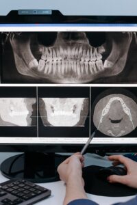
As the name suggests, panoramic X-rays provide a comprehensive view of the entire oral cavity in one image. This type of X-ray is essential in planning treatments, examining jaw and tooth relationships, or evaluating wisdom teeth problems.
- Occlusal X-rays: An Overview of Comprehensive Diagnosis
Occlusal X-rays are not as detailed as other types, but they provide a broad view of the entire arch of the teeth in either the upper or lower jaw. They play a crucial role in diagnosing issues regarding the positioning of teeth, detecting developmental abnormalities, or finding foreign objects in the mouth.
- Computerized Tomography (CT): Precision and Clarity
CT creates a 3D image of oral structures, providing unmatched detail and precision. It assists in advanced procedures like implants, root canal therapy, or diagnosing complex issues involving bones, blood vessels, and nerves.
How Does a Dental X-ray Work? The Science Behind the Screens
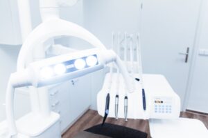
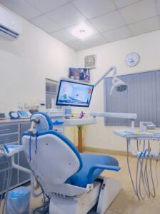
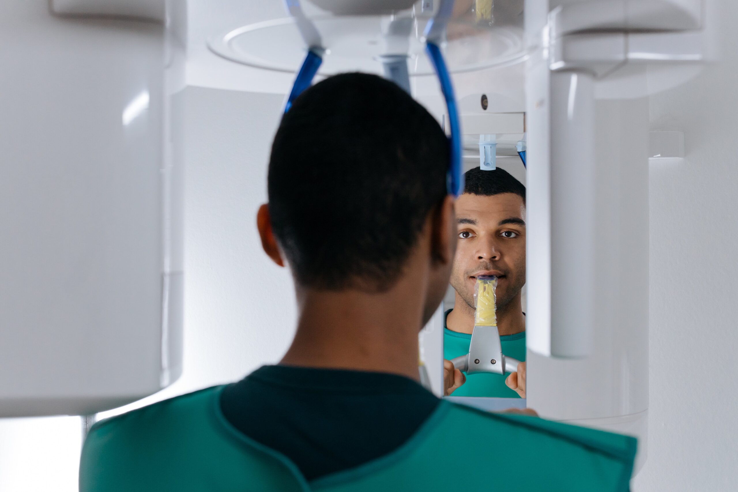
Generally, a dental X-ray procedure involves the dentist or technician covering you with a lead apron for protection, placing a small device called an X-ray film holder in your mouth, and then taking images using an X-ray machine. While detailed personal experiences may vary depending on the type of X-ray and related circumstances, the process typically works by passing radiation through oral tissues. Denser tissues such as teeth and bones absorb more radiation, appearing white on film, while softer tissues absorb less, appearing darker. Dentists decode these images to identify any abnormalities or issues.
Despite involving radiation, dental X-rays are remarkably safe. Modern technology has significantly reduced radiation exposure, making the risk negligible compared to the diagnostic benefits offered. Nevertheless, dentists always follow rigorous safety practices, which involve minimizing exposure through protective aprons and thyroid collars.
Diagnostic Benefits and Importance of Dental X-rays
Dental X-rays are essential in detecting issues invisibly lurking in your mouth. Hidden cavities, impacted teeth, jawbone damage, or even tumors can be identified in their nascent stages, making intervention timely and more effective. Moreover, X-rays also act as valuable tools in many dental procedures. Whether deciding on the approach for extraction, guiding a root canal treatment, or planning for implants, dental X-rays provide vital information contributing to the treatment’s success.
In orthodontics, X-rays play a pivotal role in determining the best course of treatment by providing a full picture of the patient’s teeth and jaws. They also allow orthodontists to monitor treatment progress and make necessary adjustments along the way.
Challenges, Risks and Controversies Around Dental X-rays
While radiation exposure from dental X-rays is relatively low, there are potential risks, especially for pregnant women or individuals highly sensitive to radiation. Regular exposure over time could theoretically lead to an increased cancer risk, emphasizing the importance of judicious, justifiable use of radiographs. Despite popular beliefs, dental X-rays don’t generate harmful levels of radiation, and precautions like lead aprons significantly reduce exposure. Moreover, they are not addictive or painful. Addressing these misconceptions is crucial in reassuring patient safety and promoting oral health.
The controversies around dental X-rays often stem from miscommunication and misinformation. While risks do exist, they are minimal, and the benefits of early and precise diagnosis far outweigh the potential drawbacks.
How to Prepare For a Dental X-ray
Your dentist will advise when you need a dental X-ray, typically during routine check-ups or when dealing with oral health problems. The frequency of X-rays depends on multiple factors, including age, current oral health status, a history of dental diseases, and any immediate symptoms or problems. Prior to the X-ray, preparation usually involves informing your dentist about any possibility of pregnancy or existing health conditions.
On the day of the X-ray, it’s advised to brush your teeth so your mouth is clean for the X-ray films. Additionally, the cost of dental X-rays may vary depending on factors such as the type of X-ray, location, insurance coverage, among others. It’s essential to discuss these aspects with your dental office and ensure you understand the costs prior to the appointment.
The Future of Dental X-rays: Trends and Advancements
Dental radiography continues to evolve with recent advancements such as digital X-rays, which reduce radiation exposure and improve image quality. Additionally, Cone Beam Computed Tomography (CBCT) has emerged, providing 3D images that are transforming dental diagnostics. The advent of Artificial Intelligence (AI) and Machine Learning (ML) holds significant promise in the field.
These technologies have the potential to automate and improve the process of detecting abnormalities, establishing diagnostics, and even predicting future oral issues from dental X-ray images. Furthermore, improving patient education and awareness about the safety, importance, and advancements in dental X-rays is crucial. This can help debunk myths, address concerns, and promote the importance of regular checks to maintain overall oral health.
In conclusion, dental X-rays are a vital aspect of oral healthcare. They serve as indispensable tools in diagnosing, treating, and various dental conditions. The recent strides in technology only promise to enhance their effectiveness and safety, revolutionizing the future of dental radiography. Despite the potential risks, when used judiciously, the diagnostic rewards certainly outweigh them. Remember, patient education, and communication is key in addressing any concerns related to dental X-rays. So, be informed, ask questions, and remember to smile for the camera – or in this case, the X-ray machine!
Our Reading dental clinic is committed to providing you with a beautiful smile every time you visit us. Whether you need x-ray or any other dental services, our team of highly trained dentists is here to provide you with the highest quality care. Our aim at Smiles in Reading is to give you a smile that is both comfortable and respectful. With online appointment scheduling, you can now receive dental care of the highest quality. Our dental professionals are here to help you enhance your smile and teeth.