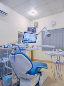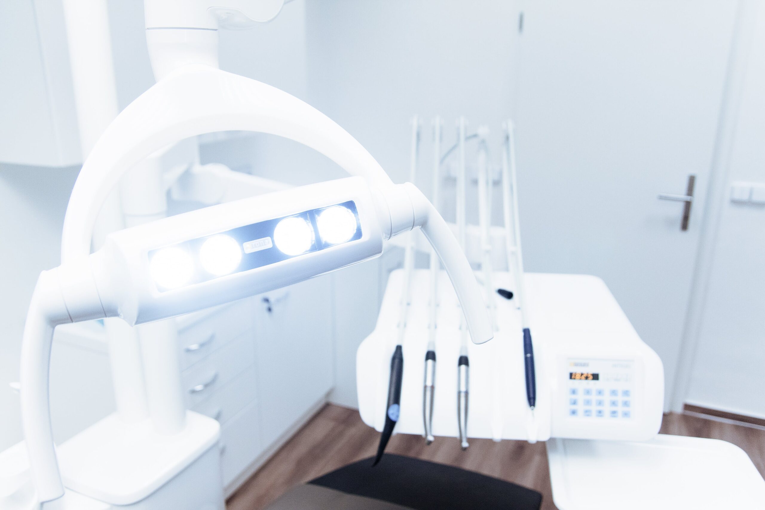
Introduction
Dental X-rays play a critical role in maintaining oral health, allowing dentists to detect and diagnose dental problems that may not be visible to the naked eye. However, it is crucial to make informed decisions about the frequency of dental X-rays to ensure both the effectiveness and safety of this diagnostic tool. In this blog post, we will explore the factors that influence dental X-ray frequency, guidelines provided by dental associations, radiation exposure, and safety measures. We will also emphasize the significance of patient-dentist communication in making informed decisions about dental X-ray frequency.
II. Factors Influencing Dental X-ray Frequency
Your oral health history, including previous dental work and existing oral health conditions, can influence the frequency of dental X-rays. For example, individuals with a history of dental fillings, root canals, or periodontal disease may require more frequent X-rays to monitor the progress of their treatment or detect any new issues. Moreover, personal habits, such as smoking or poor oral hygiene, can increase the risk of dental problems, warranting more regular X-rays. Additionally, genetic factors and family history of dental issues can also impact the recommended frequency of X-rays.
Age plays a crucial role in determining the frequency of dental X-rays. Infants and children may require X-rays more frequently to monitor the development of their teeth and identify any underlying issues, such as impacted teeth or cavities. As adolescents and young adults undergo orthodontic treatment or experience dental changes, the frequency of X-rays may increase. For adults and elderly individuals, the frequency of X-rays depends on their oral health status and risk assessment.
Certain dental issues require more frequent X-rays to ensure accurate diagnosis and effective treatment. For instance, tooth decay and cavities may necessitate X-rays to identify the extent of the damage and create a suitable treatment plan. Similarly, patients with gum disease or periodontal health issues may need X-rays to assess the condition of their gums and bone structure. Orthodontic treatment and alignment procedures often require X-rays to evaluate the progress and make necessary adjustments.
III. Guidelines for Dental X-ray Frequency
A. Recommendations from Dental Associations
- American Dental Association (ADA)
- Academy of General Dentistry (AGD)
- The International Association for Dental and Maxillofacial Radiology (IADMFR)
Several reputable dental associations provide guidelines on dental X-ray frequency. The American Dental Association (ADA) suggests that dentists evaluate each patient individually and consider their unique risk factors when deciding on the frequency of X-rays. The Academy of General Dentistry (AGD) emphasizes the importance of using X-rays as a diagnostic tool based on the patient’s overall health, oral condition, and susceptibility to dental problems. The International Association for Dental and Maxillofacial Radiology (IADMFR) also provides evidence-based recommendations to ensure safe and effective use of dental X-rays.
B. Routine Dental X-rays and Frequency
- Bitewing X-rays
- Periapical X-rays
- Panoramic X-rays
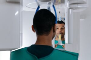
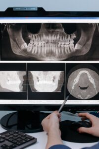
Routine dental X-rays, including bitewing, periapical, and panoramic X-rays, are commonly used to assess overall oral health. Bitewing X-rays allow dentists to detect early signs of tooth decay and monitor the progression of cavities. Periapical X-rays provide detailed images of specific teeth, including the root structure and surrounding bone, aiding in the diagnosis of dental infections or abnormalities. Panoramic X-rays capture a broad view of the entire mouth, enabling dentists to evaluate the jaw, teeth alignment, and impacted teeth.
C. Specialized X-rays and Frequency
- Cone Beam Computed Tomography (CBCT)
- Occlusal X-rays
- Extraoral Radiographs
In certain cases, specialized X-rays may be necessary to obtain more detailed information for diagnosis and treatment planning. Cone Beam Computed Tomography (CBCT) provides three-dimensional images of the teeth, jaw, and surrounding structures, offering enhanced precision. Occlusal X-rays capture a broader view of the upper and lower jaw, useful for assessing the development of teeth, identifying fractures, and locating foreign objects. Extraoral radiographs, such as panoramic X-rays, are taken from outside the mouth and provide a comprehensive view of the skull, jaw, and teeth.
IV. Understanding Radiation Exposure and Safety Measures
Radiation exposure is a concern when undergoing dental X-rays. However, it is important to understand that the radiation levels associated with dental X-rays are relatively low, especially when compared to other sources of radiation, such as natural background radiation or medical imaging procedures like CT scans. To further minimize radiation exposure, modern dental practices utilize advanced technology and techniques that significantly reduce radiation doses. Shielding measures, such as lead aprons and thyroid collars, are also employed to protect patients from unnecessary exposure.
To ensure patient safety during dental X-rays, various protective measures are implemented. Leadons and thyroid collars shield sensitive organs from radiation and reduce the risk of potential harm. Digital X-ray sensors offer advantages over film X-rays, such as lower radiation doses, immediate image availability, and better image quality. Dentists also follow proper X-ray techniques and ensure regular calibration of X-ray equipment to maintain optimal safety standards.
V. Informed Decision-Making and Patient Communication
Open communication between patients and dentists is crucial in making informed decisions about dental X-ray frequency. Patients should share any oral health concerns, changes in symptoms, and their complete medical history with the dentist to enable accurate assessments. Regular check-ups and assessments are vital not only for monitoring oral health but also for adjusting the frequency of X-rays based on the patient’s specific needs.
Moreover, maintaining the appropriate dental X-ray frequency offers several benefits in terms of oral health. Regular X-rays enable the early detection of dental issues, such as cavities or periodontal disease. Timely identification of these problems allows for prompt intervention and prevents further complications. In addition, accurate treatment planning and monitoring become possible through X-rays, ensuring that the chosen treatment approach is effective and appropriate for the patient’s specific oral condition.
In conclusion, despite the benefits, patients often have concerns and misconceptions about dental X-rays, which can hinder their decision-making process. Addressing common myths, such as excessive radiation exposure or the harmful effects of X-rays, is essential in educating patients about the actual risks and benefits associated with these diagnostic tools. By clearly explaining the importance of dental X-rays, dentists can help patients make informed decisions confidently. Remember, your dentist is your partner in oral health, and together you can make informed decisions that will benefit you in the long run.
Our Reading dental clinic is committed to providing you with a beautiful smile every time you visit us. Whether you need X-ray or any other dental services, our team of highly trained dentists is here to provide you with the highest quality care. Our aim at Smiles in Reading is to give you a smile that is both comfortable and respectful. With online appointment scheduling, you can now receive dental care of the highest quality. Our dental professionals are here to help you enhance your smile and teeth.